Instructions:
This works for dimension 3100 software 0x0531000 anyway...
Open your afm data in imageJ. Draw a line and extract a cross section (i.e. Control K). Copy and paste the data to a spreadsheet. The intensity of the image is considered a z-coordinate now. Next we need to convert the x and z columns to realspace measurements. The x conversion is easiest: open the AFM file in a text editor (e.g. notepad). Then locate the image resolution and size (for example the image is 256 pixels by 256 pixels and the scan size is 40 000 nm). Then multiply each value by 40000/256 (the x scale factor) to convert to nm (the real space units).
The Z scale is conceptually the same, but slightly more complex to do. Again we wish to make a scale factor to convert our data. to do so we must locate the following lines in the header:
\@Sens. Zscan: V 14.30724 nm/V
\@2:Z scale: V [Sens. Zscan] (0.006713765 V/LSB) 334.2918 V
Note the numbers at the end will be different in each image, they are what we need to make the image specific conversion factor. In this case the conversion is 14.30724 x 334.2918 / 65535. The first two numbers are from the file (nm per volt and volts per digital value) and the last is the number of digital levels available to the scanner. Multiply your z column by this number and you are all set.
Monday, January 9, 2012

Friday, December 30, 2011
Calibration Factors
Note these are 'pre move' measurements.
Confocal Microscope
Light Microscope
Confocal Microscope
| lens (in Air) | resolution | f (um/px) |
| 100x | 800x800 | 0.160767 |
| 100x | 1024x1024 | 0.124108 |
| 100x | 1600x1600 | 0.079653 |
| 100x | 2048x2048 | 0.063535 |
| 10x | 800x800 | 1.614726 |
| 10x | 1024x1024 | 1.015435 |
| 10x | 1600x1600 | 0.808205 |
| 10x | 2048x2048 | 0.63066 |
| lens (in water) | resolution | f (um/px) |
| 100x | 800x800 | 0.16001 |
| 100x | 1024x1024 | 0.115274 |
| 100x | 1600x1600 | 0.080559 |
Light Microscope
| um/px | |
| 50x | 0.186104 |
| 5x | 1.864454 |
Thursday, December 29, 2011
Microscope Calibrations
This post is to document how and why we calibrate the microscopes. To begin with a microscope's most basic function is to change the size of something (i.e. make it bigger) for us to view. This does not mean the object changes size, just what we see does (the image is bigger). But we may want to use the image to measure a real size or distance. For example, in the image shown below, we might want to know far it is from the bug's belly to the ruler (or the bug's leg length or width or something).

Currently we usually store an image as a digital photograph on a computer, which means the image is stored as a collection of pixels (the far right part of the image above). If I were to measure a distance I would count how many pixels from place to place. Using the example above I might say that the bug's belly is one pixel away from the ruler, at its closest point. The question is what this distance is in the 'real world', i.e. the real distance from the bug to the ruler. To say, I need to know the 'calibration factor' for my microscope.
The calibration factor is a unit conversion, usually from pixels to microns for an ordinary light microscope. To calculate the conversion factor, all we need is a known distance. In the example above, the ruler will have real distances marked on it, so we have a 'known' distance in our image. All we need to do is measure how many pixels are between two markings on the ruler, and note the dimensions of the markings. For example, say I measure 5 pixels between two millimeter marks on the ruler. My calibration factor is then given by: 1000 um / 5 pixels = 200 um / pixel. I can then take this calibration factor and convert the bug-ruler distance: 1 pixel x 200 um/pixel = 200 um. So my bug is 200 um from the ruler.

Currently we usually store an image as a digital photograph on a computer, which means the image is stored as a collection of pixels (the far right part of the image above). If I were to measure a distance I would count how many pixels from place to place. Using the example above I might say that the bug's belly is one pixel away from the ruler, at its closest point. The question is what this distance is in the 'real world', i.e. the real distance from the bug to the ruler. To say, I need to know the 'calibration factor' for my microscope.
The calibration factor is a unit conversion, usually from pixels to microns for an ordinary light microscope. To calculate the conversion factor, all we need is a known distance. In the example above, the ruler will have real distances marked on it, so we have a 'known' distance in our image. All we need to do is measure how many pixels are between two markings on the ruler, and note the dimensions of the markings. For example, say I measure 5 pixels between two millimeter marks on the ruler. My calibration factor is then given by: 1000 um / 5 pixels = 200 um / pixel. I can then take this calibration factor and convert the bug-ruler distance: 1 pixel x 200 um/pixel = 200 um. So my bug is 200 um from the ruler.
Group Photo Summer 2011
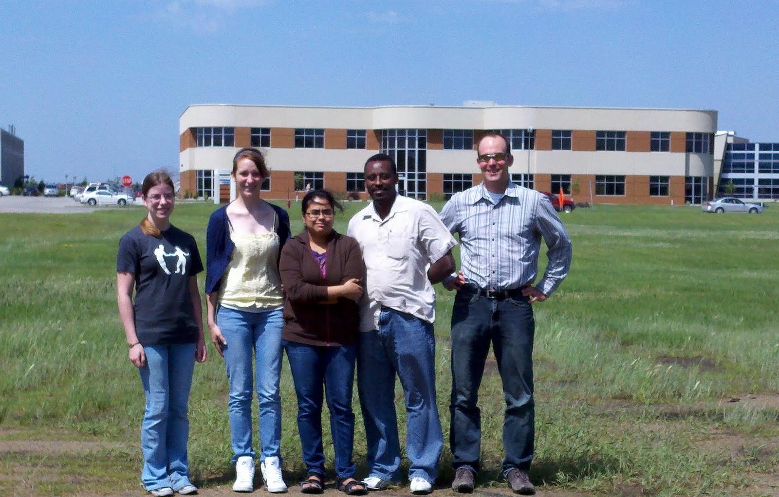
Here we are! Obviously I am a little behind on the posts!
From the left: Michelle Davis (Summer Student - Mech. Eng.), Katrina Teskey (Governor's School Student), Margaret Rozario (Summer Student - Physics), Bekele Gurmessa (Ph.D. Student - Physics), Andrew B. Croll (Boss).
Background: Batcheller Technology Center.

Subscribe to:
Posts (Atom)